Основы маммографии
On this page
- Why do I need mammograms?
- What are the main uses of mammograms?
- What do mammograms show?
- How do mammograms work?
- What are three-dimensional (3D) mammograms?
- Are mammograms safe?
A mammogram is a low-dose x-ray that allows doctors called radiologists to look for changes in breast tissue.
Why do I need mammograms?
Mammograms can be used to look for breast cancer, either as a screening test in women without symptoms or in women who have symptoms that might be from cancer. A mammogram can often find or detect breast cancer early, when it’s small and even before a lump can be felt. This is when it’s likely to be easiest to treat.
What are the main uses of mammograms?
Screening mammograms
A screening mammogram is used to look for signs of breast cancer in women who don’t have any breast symptoms or problems. X-ray pictures of each breast are taken, typically from 2 different angles.
Diagnostic mammograms
Mammograms are used to look at a woman’s breast if she has breast symptoms or if something unusual is seen on a screening mammogram. When used in this way, they are called diagnostic mammograms. They may include extra views (images) of the breast that aren’t part of screening mammograms. Sometimes diagnostic mammograms are used to screen women who were treated for breast cancer in the past.
What do mammograms show?
Mammograms can often show abnormal areas in the breast. They can’t tell for sure if an abnormal area is cancer, but they can help health care providers decide if more testing (such as a breast biopsy) is needed. The main types of breast changes found with a mammogram are:
- Calcifications
- Masses
- Asymmetries
- Distortions
Learn more about these and other breast changes in What Does the Doctor Look for on a Mammogram?
How do mammograms work?
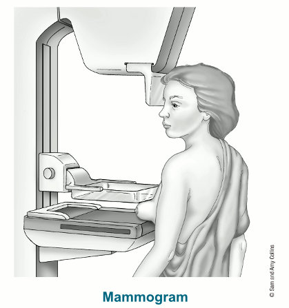
Mammograms are done with a machine designed to look only at breast tissue. The machine takes x-rays at lower doses than the x-rays done to look at other parts of the body, like the lungs or bones. The mammogram machine has 2 plates that compress or flatten the breast to spread the tissue apart. This gives a better quality picture and allows less radiation to be used.
To learn more about how they are done, see Tips for Getting a Mammogram.

In the past, mammograms were typically printed on large sheets of film. Today, digital mammograms are much more common. Digital images are recorded and saved as files in a computer.
What are three-dimensional (3D) mammograms?
Three-dimensional (3D) mammography is also known as breast tomosynthesis or digital breast tomosynthesis (DBT). As with a standard (2D) mammogram, each breast is compressed from two different angles (once from top to bottom and once from side to side) while x-rays are taken. But for a 3D mammogram, the machine takes many low-dose x-rays as it moves in a small arc around the breast. A computer then puts the images together into a series of thin slices. This allows doctors to see the breast tissues more clearly in three dimensions. (A standard two-dimensional [2D] mammogram can be taken at the same time, or it can be reconstructed from the 3D mammogram images.)
Many studies have found that 3D mammography appears to lower the chance of being called back for follow-up testing after screening. It also appears to find more breast cancers, and several studies have shown it can be helpful in women with dense breasts. A large study is now in progress to better compare outcomes between 3D mammograms and standard (2D) mammograms.
For more on 3D mammograms, see American Cancer Society Recommendations for the Early Detection of Breast Cancer.
Are mammograms safe?
Mammograms expose the breasts to small amounts of radiation. But the benefits of mammography outweigh any possible harm from the radiation exposure. Modern machines use low radiation doses to get breast x-rays that are high in image quality. On average the total dose for a typical mammogram with 2 views of each breast is about 0.4 millisieverts, or mSv. (A mSv is a measure of radiation dose.) The radiation dose from 3D mammograms can range from slightly lower to slightly higher than that from standard 2D mammograms.
To put these doses into perspective, people in the US are normally exposed to an average of about 3 mSv of radiation each year just from their natural surroundings. (This is called background radiation.) The dose of radiation used for a screening mammogram of both breasts is about the same amount of radiation a woman would get from her natural surroundings over about 7 weeks.
If there’s any chance you might be pregnant, let your health care provider and x-ray technologist know. Although the risk to the fetus is very small, and mammograms are generally thought to be safe during pregnancy, screening mammograms aren’t routinely done in pregnant women who aren't at increased risk for breast cancer.
Mammograms might also result in some women getting additional tests that don't result in a breast cancer diagnosis, but that might still have their own harms. For more on this, see Limitations of Mammograms.